The Clinical Reality of Tear Trough Fillers: A Doctor’s Guide to Risks and Safety
- RJ CLINIC
- 2 days ago
- 10 min read


The most critical decision an aesthetic doctor makes is not which product to use, but rather identifying which patients should not be treated at all. While the promise of a refreshed look is appealing, the risks of tear trough fillers are often rooted in poor patient selection or a failure to respect the complex anatomy of the lower eyelid. You may be observing a persistent shadow that makes you look fatigued, yet you are likely balancing the desire for restoration with a very real anxiety about lumps or vascular complications.
We believe that clinical safety and aesthetic subtlety must always coexist. This guide provides a nuanced look at how a meticulous anatomical assessment prevents long term issues like the Tyndall effect or chronic swelling. You will learn the vital distinction between true volume loss and fat pad protrusion, helping you determine if this treatment is the right path for your specific anatomy. By prioritizing medical integrity over a one size fits all approach, we ensure your journey toward a restored, natural appearance is both predictable and secure.
Key Takeaways
Understand why the lower eyelid's thin skin requires specific product selection to prevent visible lumps or surface irregularities.
Identify the clinical risks of tear trough fillers, including the Tyndall effect, and how expert depth of injection mitigates these concerns.
Learn how to perform a simple 'snap test' at home to assess if your skin laxity is suitable for a filler treatment.
Explore why Dr Renee and Dr Cheok prioritize blunt-tip cannulas to preserve vascular integrity and minimize downtime during the procedure.
Table of Contents
Beyond the 'Tired Look': Why Tear Trough Fillers Are Anatomically Challenging
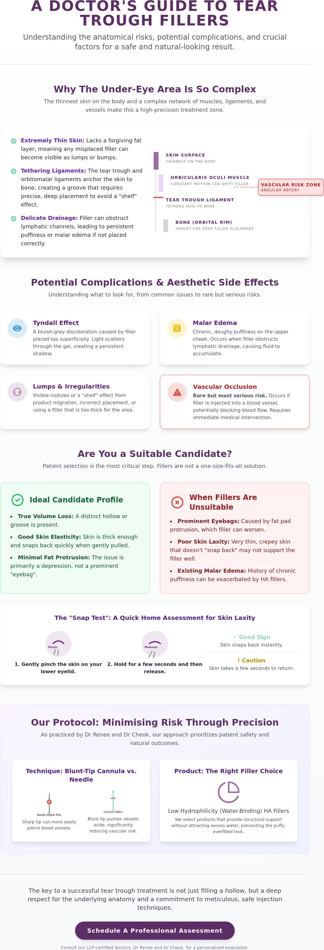
The lower eyelid presents a unique challenge because it possesses the thinnest skin on the human body. This lack of dermal thickness means that even the slightest misplacement of an Injectable filler overview can result in visible lumps or an unnatural contour. Unlike other areas of the face where a layer of subcutaneous fat provides a buffer, the periocular region requires a practitioner to work in a space where the margin for error is measured in millimetres. In clinical practice, the risks of tear trough fillers are best understood as a convergence of intricate periocular anatomy and the inherent hydrophilicity of the chosen injectable product.
The tear trough itself is not merely a hollow caused by fat loss; it's often defined by the tear trough ligament. This structure tethers the skin to the underlying bone, creating a fixed groove. If a doctor simply fills this groove without understanding the tension of the ligament, the product may migrate or create a shelf effect above the tether point. Additionally, the proximity of the lymphatic drainage system is a frequent cause of post-procedure dissatisfaction. If filler is placed too superficially or in excessive volumes, it can obstruct lymphatic flow, leading to chronic malar oedema or persistent morning puffiness that doesn't resolve with time.
The Role of the Periocular Anatomy
The orbitomalar ligament is the primary architect of the hollow appearance. It creates a physical barrier that divides the eyelid from the cheek. When we inject in this zone, we're often placing material beneath the orbicularis oculi muscle. This muscle is constantly in motion. Every blink or squint can shift poorly integrated filler. Because this is a high-risk zone, vascular mapping is a non-negotiable part of our protocol. We must account for the angular artery and the infraorbital network to avoid vascular compromise, which remains one of the most serious risks of tear trough fillers.
Hydrophilic Nature of Fillers
Most dermal fillers are composed of Hyaluronic Acid (HA), a molecule that naturally attracts water. While this is beneficial for lip volume, it can be problematic under the eyes. High hydrophilicity leads to tissue saturation rather than structural lifting. This often results in the puffy look that many patients fear. We prioritise fillers with low water-binding capacity to ensure the result remains crisp and stable. The goal is to provide subtle support to the anatomy without causing the tissue to hold excess fluid, which often fluctuates based on your salt intake or sleep quality.
Managing Common and Rare Complications: From Bruising to Vascular Safety
Distinguishing between expected recovery and genuine medical complications is the first step toward a safe aesthetic journey. While minor bruising and transient swelling are typical in the first 72 hours, persistent issues require clinical intervention. One of the most frequently discussed risks of tear trough fillers is the Tyndall effect, where the under-eye area takes on a faint blue or grey hue. This occurs when filler is injected too close to the surface, causing light to scatter through the hyaluronic acid molecule rather than reflecting naturally.
Malar oedema represents another significant concern, manifesting as chronic, doughy swelling on the upper cheek. This happens when the filler inadvertently obstructs the delicate lymphatic channels, preventing fluid from draining efficiently. Unlike standard post-procedural swelling, malar oedema often fluctuates throughout the day and may persist for months if the product isn't strategically dissolved. Vascular occlusion in the infraorbital artery is mitigated by using micro-cannulas rather than needles, as these blunt-tipped tools are significantly less likely to penetrate vessel walls. According to the FDA guide to dermal filler risks, the most serious complications involve accidental intravascular injection, which underscores why anatomical expertise is non-negotiable.
Aesthetic Side Effects: Lumps and Discolouration
Lumps often arise from product migration or superficial placement within the orbicularis oculi muscle. When these aesthetic errors occur, we utilize hyaluronidase, a specialized enzyme that safely breaks down the filler. This allows us to reset the canvas and correct any asymmetry or visible "shelving" of the product. If you are currently experiencing persistent puffiness or visible product from a previous treatment, an expert assessment for eyebag treatment or filler reversal can help restore a natural contour.
Severe Medical Risks and Mitigation
Delayed onset nodules or granulomas may appear months after treatment as a late-stage immune response to the foreign material. Maintaining a sterile clinical environment is paramount to preventing biofilm formation and long-term infection. Our safety protocol dictates that a comprehensive emergency 'dissolving kit' is always on standby. This ensures that any sign of vascular distress or delayed inflammatory response can be addressed with immediate, high-dose enzymatic treatment to protect the integrity of the surrounding tissue.
The 'Candidate Filter': When Fillers Are the Wrong Solution
Determining suitability is the most overlooked step in the patient journey. Many individuals seek dermal fillers to camouflage "dark circles" or "hollows" without realizing that their underlying anatomy may actually contraindicate the procedure. Understanding the risks of tear trough fillers involves recognizing when the anatomy requires a different modality entirely. If we ignore these anatomical signals, the result is rarely a refreshed appearance; instead, it often leads to a heavy, overfilled look that distorts the natural mid-face transition.
The "Snap Test" is a fundamental clinical assessment we use to evaluate lower lid laxity. By gently pulling the lower eyelid skin down and observing how quickly it returns to its original position, we can determine if the skin has the structural integrity to support Hyaluronic Acid. If the skin is too lax or "crepy," the filler will likely sit as a visible ridge rather than integrating into the tissue. A comprehensive review of tear trough filler complications highlights that poor patient selection is a primary driver of long term dissatisfaction and chronic swelling. When skin quality is compromised, adding volume often exacerbates the problem by weighing down the delicate periocular structures.
Identifying Poor Candidates
True eyebags, known clinically as fat prolapse, cannot be effectively "fixed" with filler. Attempting to fill the groove beneath a protruding fat pad often creates a "pillow face" effect, where the entire under-eye area looks unnaturally flat and water-logged. This is particularly risky for patients prone to seasonal allergies or fluid retention, as HA fillers can further obstruct already sluggish lymphatic drainage. Patients with festoons; malar mounds that sit on the cheekbone; are also poor candidates, as fillers in this region almost always lead to chronic, visible puffiness.
Clinical Alternatives for Eye Rejuvenation
When fillers are deemed unsuitable, we pivot to treatments that address the root cause of the concern. For those with significant fat protrusion, laser eyebag removal offers a way to reduce fat pads without the risks of tear trough fillers. In cases where structural sagging is the primary issue, eyebag surgery provides a permanent correction that no injectable can replicate. We often find that combining skin tightening with a conservative approach to volume yields a far more sophisticated and organic-looking result. If you are unsure whether your anatomy is right for fillers, a professional assessment for eyebag treatment can provide the clarity you need to move forward safely.
The RJ Clinic Protocol: Minimising Risk Through Precision
Safety in the periocular region isn't an accident; it's the result of a disciplined clinical protocol. At RJ Clinic, we recognise that the risks of tear trough fillers are significantly reduced when the procedure is performed by practitioners who possess a deep understanding of facial layers. Dr Renee and Dr Cheok are LCP-certified doctors, a credential that signifies they've met the rigorous standards set by the Ministry of Health Malaysia for aesthetic practice. This certification is your assurance that your treatment is guided by medical expertise rather than mere technical application.
One of the cornerstone safety measures we employ is the use of blunt-tip cannulas. Unlike traditional sharp needles, which can easily pierce blood vessels, a cannula's rounded end allows it to glide through the tissue. This choice is vital for preserving vascular integrity and minimising the risks of tear trough fillers associated with accidental intravascular injection. We also adhere to a 'less is more' philosophy. It's far safer to under-correct and add a touch more product later than to over-fill the area, which often leads to the unnatural, puffy appearance that many patients fear.
The Consultation and Mapping Process
Every successful outcome begins with a comprehensive facial assessment. We don't just look at the hollow; we evaluate the mid-face and cheek support first. Often, adding volume to the cheek provides the necessary lift to improve the tear trough without needing direct injection into the delicate eyelid skin. Choosing the right product is equally critical. We select specifically formulated, low-hydrophilic HA fillers that don't attract excessive water, ensuring your results remain stable and crisp even months after the procedure. We also take the time to manage expectations, explaining where volume can help and where laser treatments might be better for dark circles.
Safety Standards in Kuala Lumpur
In the Malaysian aesthetic landscape, LCP certification acts as a primary trust signal, distinguishing medical professionals from unlicensed providers. We exclusively use FDA-approved, premium injectables to ensure biocompatibility and predictable degradation. Following your session, we provide a clear monitoring protocol for the first 48 hours, focusing on any signs of unusual pain or skin colour changes. If you're ready for a safer, medically-led approach to restoration, we invite you to schedule a detailed anatomical assessment for eye rejuvenation with our specialists.
Securing Your Aesthetic Future Through Clinical Precision
Achieving a refreshed lower eyelid requires more than just product; it demands a deep respect for the intricate anatomy of the periocular region. By understanding the risks of tear trough fillers and the necessity of a rigorous candidate filter, you can avoid common pitfalls like chronic swelling or unnatural contours. True restoration is found in the balance between clinical precision and artistic subtlety, ensuring that every intervention enhances your natural features without compromise.
Our approach at RJ Clinic centers on this meticulous methodology. Dr Renee and Dr Cheok, our LCP-certified aesthetic physicians, specialize in micro-cannula techniques designed to protect vascular integrity while delivering refined results. We don't believe in one size fits all solutions; instead, we focus on a subtle, natural-looking eye restoration that respects your unique structural needs. Whether your anatomy requires a conservative filler approach or an alternative modality, we prioritize your safety above all else.
If you're ready to explore a personalized treatment plan backed by medical integrity, we invite you to Book a Consultation with our LCP-certified doctors at RJ Clinic. We look forward to helping you regain your confidence through a secure and sophisticated aesthetic journey.
Frequently Asked Questions
Can tear trough filler cause permanent blindness?
Permanent blindness is an extremely rare but documented risk of tear trough fillers, occurring when filler is accidentally injected into a blood vessel that supplies the retina. While the incidence is very low, this complication underscores why we use blunt-tip cannulas and meticulous anatomical mapping. At RJ Clinic, our LCP-certified doctors follow strict safety protocols to identify and avoid high-risk vascular structures throughout the procedure.
How do I know if my filler has migrated or if it is just swelling?
Standard swelling typically resolves within seven to fourteen days; migration, however, presents as a persistent, unnatural bulge or "shelf" that often appears outside the original treatment area months later. If you notice the filler shifting toward the cheek or creating a ridge when you smile, it's likely migrated. This is usually the result of over-filling or placing product into the wrong anatomical plane.
Is the Tyndall effect permanent if left untreated?
The Tyndall effect is not permanent, but it will not resolve on its own without medical intervention. This bluish discoloration happens when hyaluronic acid is placed too superficially, causing light to scatter through the skin. To correct this, we use hyaluronidase to safely dissolve the misplaced product. Once the filler is broken down, the blue tint disappears almost immediately, restoring your natural skin tone.
What should I do if I feel a hard lump under my eye after filler?
If you detect a hard lump under the eye, you should schedule a clinical review rather than attempting to massage the area yourself. Lumps can be caused by localized product accumulation, bruising, or, in some cases, a delayed inflammatory nodule. Dr Renee or Dr Cheok will assess the lump to determine if it requires enzymatic dissolving or if it is simply a transient part of the healing process.
Why do my eyes look puffier in the morning after getting fillers?
Morning puffiness occurs because hyaluronic acid is naturally hydrophilic, meaning it attracts and holds water. When you lie flat at night, fluid accumulates in the facial tissues, and the filler can exacerbate this by slightly obstructing lymphatic drainage. This usually settles once you are upright and moving; however, if the puffiness is severe or persistent, it may indicate that the filler volume is too high for your anatomy.
How long do the risks of tear trough fillers last after the procedure?
While acute risks like vascular occlusion or infection appear almost immediately, other risks of tear trough fillers, such as malar oedema or migration, can manifest months or even years after the initial treatment. Hyaluronic acid is long-lasting in the periocular area because there is little tissue movement to break it down. Continuous monitoring and choosing a practitioner with long-term follow-up protocols are essential for managing these late-onset complications.


Comments